Abdominal Anatomy : Anatomy Of The Female Abdomen And Pelvis Cut Away View Doctor Stock - Identify some abdominal pathology on medical images.
byAdmin-
0
Abdominal Anatomy : Anatomy Of The Female Abdomen And Pelvis Cut Away View Doctor Stock - Identify some abdominal pathology on medical images.. Understanding abdominal anatomy and physiology is essential to understanding the human body as a whole. The abdomen (colloquially called the belly, tummy, midriff or stomach) is the part of the body between the thorax (chest) and pelvis, in humans and in other vertebrates. Two layers in abdomenfatty superficial layer (camper's fascia)deeper membranous layer (scarper's fascia). Laterally by the midaxillary line; Abdominal surface anatomy can be described when viewed from in front of the abdomen in 2 ways:
A good amount of area is covered by the abdominal wall. The abdominal divisions should be used in conjunction with other diagnostic approaches in order to accurately diagnose a patient's condition. The xiphoid process and costal. Transversus abdominis muscle internal abdominal oblique muscle rectus abdominis muscle external abdominal oblique muscle pyramidalis muscle. Mr anatomy of the abdominal wall demonstrating the three flat muscles (short arrow);
Abdominal Surface Anatomy Creative Commons Illustration Radiology Case Radiopaedia Org from prod-images-static.radiopaedia.org The xiphoid process and costal. Laterally by the midaxillary line; • abdominal wall • upper gi tract • lower gi tract • kidneys and retroperitoneum • inguinal region. Windham was previously a surgical oncologist in the sarcoma program of the h. Radiology basics of abdominal ct anatomy with annotated coronal images and scrollable axial images to help medical students and junior doctors learning anatomy. The transversus abdominis muscle is the deepest of the abdominal muscles, lying internally to the internal abdominal obliques. The epigastric vessels (long arrow); Its upper boundary is the diaphragm, a sheet of muscle and connective tissue that separates it the abdominal organs are supported and protected by the bones of the pelvis and ribcage and are covered by the greater omentum, a fold of peritoneum.
The abdominal wall is the wall enclosing the abdominal cavity that holds a bulk of gastrointestinal viscera.
Identify abdominal anatomical structures in a variety of medical imaging platforms. We're going to take apart a plastic anatomy model and see what we can find in the abdomen. The linea semilunaris (open arrow); 5 name the nine abdominal regions and their main contents. Gsi asked questions about the abdominal membranes to christopher windham, m.d. A collection of articles covering abdominal anatomy, including abdominal wall anatomy and abdominal cavity anatomy. Radiology basics of abdominal ct anatomy with annotated coronal images and scrollable axial images to help medical students and junior doctors learning anatomy. Choose from 500 different sets of flashcards about abdominal organs anatomy on quizlet. Abdominal anatomy seen on ct. Who better to review abdominal anatomy with, than an experienced expert? Its upper boundary is the diaphragm, a sheet of muscle and connective tissue that separates it the abdominal organs are supported and protected by the bones of the pelvis and ribcage and are covered by the greater omentum, a fold of peritoneum. Identify some abdominal pathology on medical images. Abdominal cavity, largest hollow space of the body.
The linea semilunaris (open arrow); Laterally by the midaxillary line; Its upper boundary is the diaphragm, a sheet of muscle and connective tissue that separates it the abdominal organs are supported and protected by the bones of the pelvis and ribcage and are covered by the greater omentum, a fold of peritoneum. 5 name the nine abdominal regions and their main contents. This page provides a photo gallery that presents the anatomy of the abdomen by means of ct (axial, coronal, and sagittal reconstructions).
Medically Accurate Illustration Of Abdominal Anatomy Stock Photo Picture And Royalty Free Image Image 44208566 from previews.123rf.com Therefore, a firm grasp of abdominal anatomy is necessary to effectively diagnose and treat patients. Identify some abdominal pathology on medical images. The linea alba (open arrowhead); The abdominal divisions should be used in conjunction with other diagnostic approaches in order to accurately diagnose a patient's condition. The quadratus lumborum muscle (black arrow). We're going to take apart a plastic anatomy model and see what we can find in the abdomen. This muscle forms the anterior and lateral abdominal wall. But with the use of smart technology, you can learn faster and master abdomen anatomy in no time!
The abdominal divisions should be used in conjunction with other diagnostic approaches in order to accurately diagnose a patient's condition.
The abdominal divisions should be used in conjunction with other diagnostic approaches in order to accurately diagnose a patient's condition. • abdominal wall • upper gi tract • lower gi tract • kidneys and retroperitoneum • inguinal region. A collection of anatomy notes covering the key anatomy concepts that medical students need to learn. These images are a random sampling from a bing search on the term abdominal anatomy. click on the image (or right click) to open the source website in a new browser window. The transversus abdominis muscle is the deepest of the abdominal muscles, lying internally to the internal abdominal obliques. Become familiar with the anatomical divisions by exploring the world's most advanced 3d anatomy platform in complete anatomy. Understanding abdominal anatomy and physiology is essential to understanding the human body as a whole. Most students entering ultrasound have some basic understanding of anatomy. Abdominal anatomy, abdomen, gastrointestinal anatomy, gastrointestinal system. Its upper boundary is the diaphragm, a sheet of muscle and connective tissue that separates it the abdominal organs are supported and protected by the bones of the pelvis and ribcage and are covered by the greater omentum, a fold of peritoneum. Divided into 9 regions by two vertical and two horizontal imaginary planes. Two layers in abdomenfatty superficial layer (camper's fascia)deeper membranous layer (scarper's fascia). Abdominal anatomy seen on ct.
Identify some abdominal pathology on medical images. Compare and contrast the different medical imaging modalities presented in the tutorials. We're going to take apart a plastic anatomy model and see what we can find in the abdomen. You will learn the anatomical basis of pain and how to apply this knowledge in the diagnostic process. Lee moffitt cancer center & research institute in.
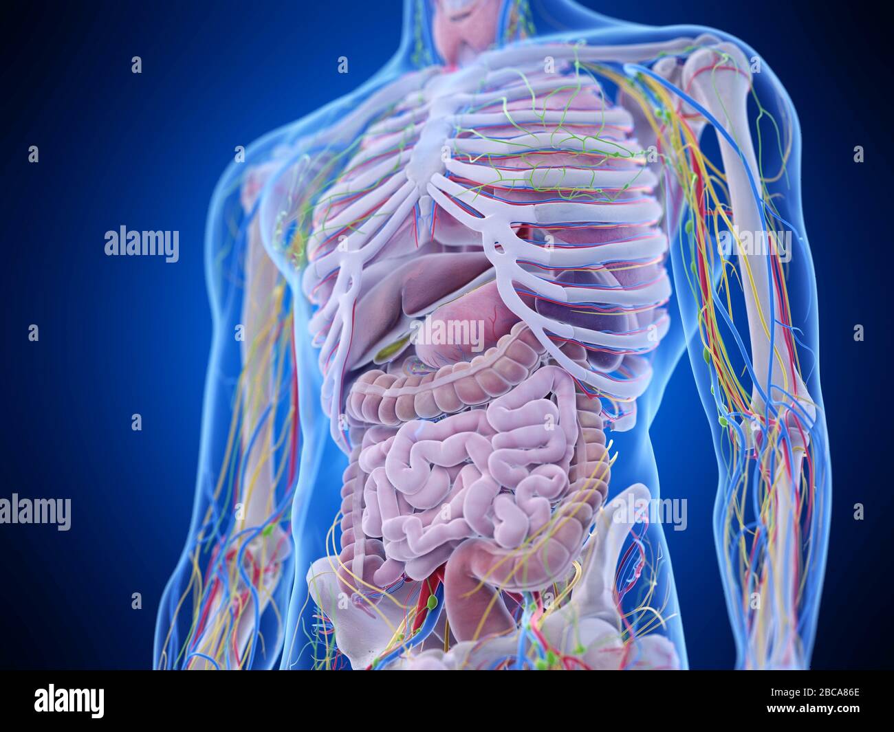
Abdominal Anatomy Illustration Stock Photo Alamy from c8.alamy.com Understanding abdominal anatomy and physiology is essential to understanding the human body as a whole. The linea alba (open arrowhead); These images are a random sampling from a bing search on the term abdominal anatomy. click on the image (or right click) to open the source website in a new browser window. The abdominal divisions should be used in conjunction with other diagnostic approaches in order to accurately diagnose a patient's condition. Become familiar with the anatomical divisions by exploring the world's most advanced 3d anatomy platform in complete anatomy. The linea semilunaris (open arrow); Mr anatomy of the abdominal wall demonstrating the three flat muscles (short arrow); Radiology basics of abdominal ct anatomy with annotated coronal images and scrollable axial images to help medical students and junior doctors learning anatomy.
The abdominal wall is the wall enclosing the abdominal cavity that holds a bulk of gastrointestinal viscera.
Understanding abdominal anatomy and physiology is essential to understanding the human body as a whole. 5 name the nine abdominal regions and their main contents. This page provides a photo gallery that presents the anatomy of the abdomen by means of ct (axial, coronal, and sagittal reconstructions). And inferiorly by the symphysis pubis, pubic tubercle, inguinal ligament, anterior superior iliac spine, and. Become familiar with the anatomical divisions by exploring the world's most advanced 3d anatomy platform in complete anatomy. Learn about abdominal organs anatomy with free interactive flashcards. The quadratus lumborum muscle (black arrow). Anatomy of the abdominal wall, inguinal region & hernias, hernias, gut and peritoneal cavity. The above lines intersect and divide the abdomen into nine regions (clockwise from the top) The epigastric vessels (long arrow); A good amount of area is covered by the abdominal wall. Windham was previously a surgical oncologist in the sarcoma program of the h. The linea alba (open arrowhead);